Please wait a minute...
欢迎访问《电化学(中英文)》期刊官方网站,今天是
图/表 详细信息
金电极表面有序分子膜的电化学原位偏振调制红外反射光谱研究
苏章菲, 陈爱成, Jacek Lipkowski
电化学(中英文)    2025, 31 (6): 2417003-.   DOI:10.61558/2993-074X.3528
摘要   (4 HTML2 PDF(pc) (3269KB)(167)  

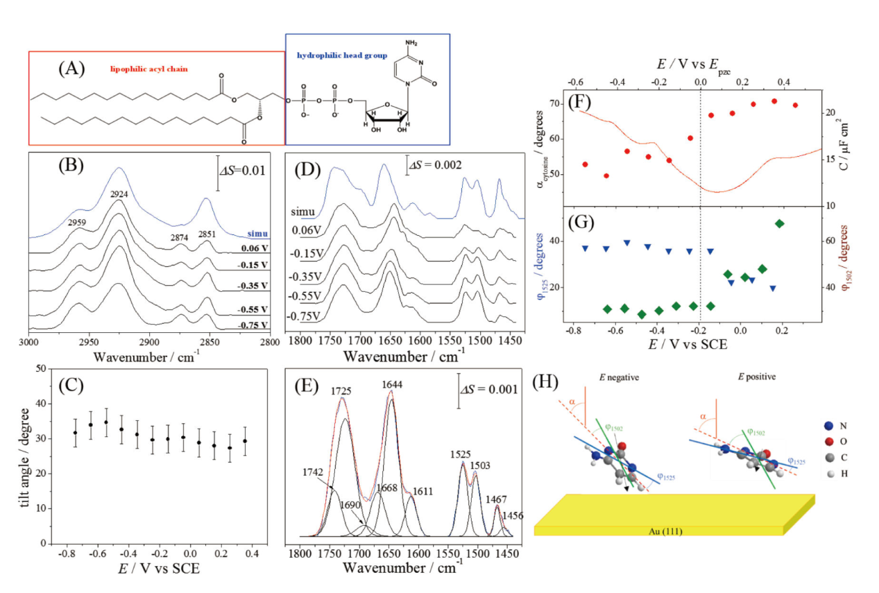
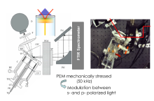
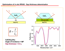
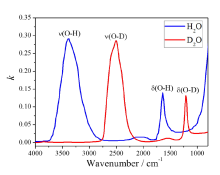
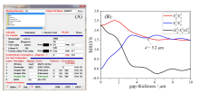
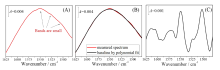
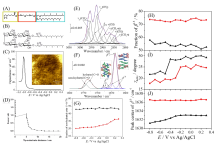
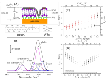
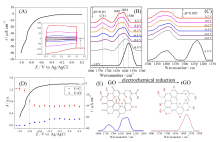
本综述探讨了电化学原位偏振调制红外反射吸收光谱在电极表面薄膜结构、取向和构象研究中的应用。该技术基于红外光谱表面选律,利用p偏振光在金属表面的增强和s偏振光的衰减特性,通过两者的差谱消除溶剂背景吸收,从而获取单一电极电位下表面物种的红外吸收信息。相比之下,另外两种流行的原位红外光谱技术,差减归一化界面傅立叶变换红外光谱和表面增强红外吸收光谱,需要进行电位差谱以消除本体溶液的信号。本文首先简要介绍了偏振调制红外反射吸收光谱的操作流程及消除背景吸收的方法,随后通过三个实例展示了该技术在仿生生物膜研究中的应用:束缚磷脂双层膜、大肠杆菌素在磷脂双层中的结构分析,以及金电极表面核脂单层膜的研究。最后,以氧化石墨烯在电化学还原过程中的结构变化为例,阐述了偏振调制红外反射吸收光谱在材料科学中的广阔应用前景。


View image in article
Figure 9. (A) Molecular structure of 1,2-dipalmitoyl-sn-glycero-3-cytidine nucleolipid. (B) PM IRRAS spectra of the nucleolipid monolayer in the C-H stretching region at selected potentials. Reprinted with permission from Ref. [36]. Copyright 2019 American Chemical Society. (C) The average tilt angle of the trans fragments of the acyl chains in the nucleolipid monolayer. Reprinted with permission from Ref. [36]. Copyright 2019 American Chemical Society. (D) PM IRRAS spectra of the nucleolipid monolayer at the Au(111) surface in the cytidine spectral region at selected electrode potentials, and (E) deconvolution of the average PM IRRAS spectrum in the cytidine spectral region. Reprinted with permission from Ref. [36]. Copyright 2019 American Chemical Society. (F) Tilt angle of the molecular plane (solid circles) and differential capacitance curve. Reprinted with permission from Ref. [36]. Copyright 2019 American Chemical Society. (G) Angles between the projection of surface normal on the molecular plane and the transition dipole directions of the vibrations at 1502 cm-1 (dark green diamonds) and 1525 cm-1 (blue triangles). Reprinted with permission from Ref. [36]. Copyright 2019 American Chemical Society. (H) Qualitative picture of the cytosine moiety orientation at negative and positive potentials. The lines represent the directions of the electrode surface normal (the solid red line), the projected surface normal on the molecular plane (the dashed red line), the transition dipole of the vibration at 1502 cm-1 (the green solid line) and the transition dipole of the vibration at 1523 cm-1 (the blue solid line). The black arrow represents the permanent dipole of the cytosine moiety. Reprinted with permission from Ref. [36]. Copyright 2019 American Chemical Society.
本文的其它图/表