金电极表面有序分子膜的电化学原位偏振调制红外反射光谱研究
PM IRRAS Studies of Organized Molecular Films at a Gold Electrode Surface
金电极表面有序分子膜的电化学原位偏振调制红外反射光谱研究 |
| 苏章菲, 陈爱成, Jacek Lipkowski |
|
PM IRRAS Studies of Organized Molecular Films at a Gold Electrode Surface |
| Zhang-Fei Su, Ai-Cheng Chen, Jacek Lipkowski |
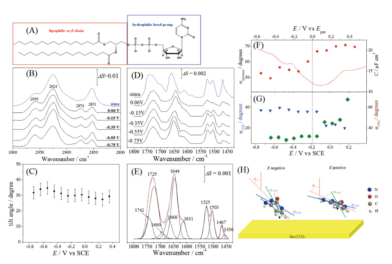
| Figure 9. (A) Molecular structure of 1,2-dipalmitoyl-sn-glycero-3-cytidine nucleolipid. (B) PM IRRAS spectra of the nucleolipid monolayer in the C-H stretching region at selected potentials. Reprinted with permission from Ref. [36]. Copyright 2019 American Chemical Society. (C) The average tilt angle of the trans fragments of the acyl chains in the nucleolipid monolayer. Reprinted with permission from Ref. [36]. Copyright 2019 American Chemical Society. (D) PM IRRAS spectra of the nucleolipid monolayer at the Au(111) surface in the cytidine spectral region at selected electrode potentials, and (E) deconvolution of the average PM IRRAS spectrum in the cytidine spectral region. Reprinted with permission from Ref. [36]. Copyright 2019 American Chemical Society. (F) Tilt angle of the molecular plane (solid circles) and differential capacitance curve. Reprinted with permission from Ref. [36]. Copyright 2019 American Chemical Society. (G) Angles between the projection of surface normal on the molecular plane and the transition dipole directions of the vibrations at 1502 cm-1 (dark green diamonds) and 1525 cm-1 (blue triangles). Reprinted with permission from Ref. [36]. Copyright 2019 American Chemical Society. (H) Qualitative picture of the cytosine moiety orientation at negative and positive potentials. The lines represent the directions of the electrode surface normal (the solid red line), the projected surface normal on the molecular plane (the dashed red line), the transition dipole of the vibration at 1502 cm-1 (the green solid line) and the transition dipole of the vibration at 1523 cm-1 (the blue solid line). The black arrow represents the permanent dipole of the cytosine moiety. Reprinted with permission from Ref. [36]. Copyright 2019 American Chemical Society. |
|
|