欢迎访问《电化学(中英文)》期刊官方网站,今天是
研究论文

氢气气泡模板电化学诱导沉积纳-微米二级结构钙磷盐生物材料的研究

  • 王卉 ,
  • 林昌健 ,
  • 胡仁 ,
  • 张克勤 ,
  • 段红平 ,
  • 董镶
展开
  • 1. 苏州大学 现代丝绸国家工程实验室,纺织与服装工程学院,江苏 苏州 215123; 2. 厦门大学 固体表面物理化学国家重点实验室,化学化工学院化学系,福建 厦门 361005; 3. 北京技术工程研究中心,北京纳通科技集团有限公司,北京 100082

收稿日期: 2013-02-28

  修回日期: 2013-04-08

  网络出版日期: 2013-04-15

基金资助

国家科技支撑计划(No. 2012BAI07B09)国家自然科学基金项目(No. 51203108),江苏省自然科学基金项目(No. BK2011355)和江苏省高校自然科学研究项目(No. 11KJB430011)资助

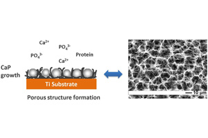
Study on Hydrogen Bubble Template Fabrication of Porous Biomaterials Coatings by Electrochemically Induced Deposition

  • WANG Hui ,
  • LIN Chang-Jian ,
  • HU Ren ,
  • ZHANG Ke-Qin ,
  • DUAN Hong-Ping ,
  • DONG Xiang
Expand
  • 1. National Engineering Laboratory for Modern Silk, College of Textile and Clothing Engineering, Soochow University, Suzhou 215123, China; 2. State Key Lab of Physical Chemistry of Solid Surfaces, Department of Chemistry, College of Chemistry and Chemical Engineering, Xiamen University, Xiamen 361005, Fujian, China; 3. Beijing Engineering Research Center, Beijing Naton Medical Group, Beijing 100082, China

Received date: 2013-02-28

  Revised date: 2013-04-08

  Online published: 2013-04-15

摘要

生物材料的多孔结构对于植入后细胞的响应及其与机体组织的有效整合有着决定性的影响. 采用电化学沉积方法在钛基表面成功制备多孔钙磷盐及钙磷盐/蛋白质复合膜层. 本文选择合适的电解液浓度、温度、电流密度、时间和蛋白质添加剂等,可有效地控制钙磷盐晶体的形状、尺寸和柔韧性,并初步探讨了氢气气泡模板的作用机制. 研究结果表明,动态氢气气泡是一种有效的模板,可控制钙磷盐晶体的生长速度,成功构筑纳-微米二级结构钙磷盐生物材料.

本文引用格式

王卉 , 林昌健 , 胡仁 , 张克勤 , 段红平 , 董镶 . 氢气气泡模板电化学诱导沉积纳-微米二级结构钙磷盐生物材料的研究[J]. 电化学, 2013 , 19(6) : 501 -506 . DOI: 10.13208/j.electrochem.130216

Abstract

So far, the pore architecture in biomaterials plays a critical role on the cell response and integration between the biomaterials and implanted environment. In this study, porous calcium phosphate (CaP) coatings and CaP/protein composite coatings have been successfully constructed on titanium substrate by using an electrochemically induced deposition technique. The shape, size and pliability of CaP crystals are controlled by electrolyte concentration, temperature, current density, time and protein additive in preparing process. In addition, the formation mechanism of the porous structure is discussed based on the “hydrogen bubble template” model. It demonstrates that the growth velocity of CaP crystals should match well with the forming-disappearing velocity of hydrogen bubble, and the pliability of the CaP crystals should fit with soft bubble. As a result, dynamic hydrogen bubble can act as an effective template to construct the nano-micro porous structured biomaterials coatings by controlling the growth velocity of CaP crystals.

参考文献

[1]Liu X Y, Paul K C, Ding C X. Surface modification of titanium, titanium alloys, and related materials for biomedical applications[J]. Materials Science and Engineering: R: Reports, 2004, 47(3/4): 49-121.
[2]Puleo D A, Nanci A. Understanding and controlling the bone-implant interface[J]. Biomaterials, 1999, 20: 2311-2321.
Schuler M, Trentin D, Textor M, et al. Biomedical interfaces: Titanium surface technology for implants and cell carriers[J]. Nanomedicine, 2006, 1(4): 449-463.
[3]de Jonge L T, Leeuwenburgh S C G, Wolke J G C, et al. Organic-inorganic surface modifications for titanium implant surfaces[J]. Pharmaceutical Research, 2008, 25(10): 2357-2369.
[4]Cheng X, Filiaggi M, Roscoe S G. Electrochemically assisted co-precipitation of protein with calcium phosphate coatings on titanium alloy[J]. Biomaterials, 2004, 25(23): 5395-5403.
[5]Fan Y, Duan K, Wang R. A composite coating by electrolysis-induced collagen self-assembly and calcium phosphate mineralization[J]. Biomaterials, 2005, 26(14): 1623-1632.
[6]Wang H, Lin C J, Hu R, et al. A novel nano-micro structured octacalcium phosphate/protein composite coating on titanium by using an electrochemically induced deposition[J]. Journal of Biomedical Materials Research Part A, 2008, 87(3): 698-705.
[7]Ren H, Lin C J, Shi H Y, et al. Electrochemical deposition mechanism of calcium phosphate coating in dilute Ca-P electrolyte system[J]. Materials Chemistry and Physics, 2009, 115(2/3): 718-723.
[8]Zhang Y F, Fan W, Ma Z C, et al. The effects of pore architecture in silk fibroin scaffolds on the growth and differentiation of mesenchymal stem cells expressing BMP7[J]. Acta Biomaterialia, 2010, 6(8): 3021-3028.
[9]Yan L P, Oliveira J M, Oliveira A L, et al. Macro/microporous silk fibroin scaffolds with potential for articular cartilage and meniscus tissue engineering applications[J]. Acta Biomaterialia, 2012, 8(1): 289-301.
[10]Mandal B B, Kundu S C. Cell proliferation and migration in silk fibroin 3D scaffolds[J]. Biomaterials, 2009, 3(15), 2956-2965.
[11]Sun W, Puzas J E, Sheu T J, et al. Nano- to microscale porous silicon as a cell interface for bone-tissue engineering[J]. Advanced Materials, 2007, 19(7): 921-924.
[12]Fowler B O, Markovic M, Brown W E. Octacalcium phosphate. 3. Infrared and Raman vibrational spectra[J]. Chemistry of Materials, 1993, 5(10): 1417-1423.
[13]Narasaraju T S B, Phebe D E. Some physico-chemical aspects of hydroxyapatite[J]. Journal of Materials Science, 1996, 31(1): 1-21.
[14]Fan Y W, Wang R Z. Submicrometer-sized vaterite tubes formed through nanobubble-templated crystal growth[J]. Advanced Materials, 2005, 17(19): 2384-2388.
[15]Li Y, Song Y Y, Yang C, et al. Hydrogen bubble dynamic template synthesis of porous gold for nonenzymatic electrochemical detection of glucose[J]. Electrochemistry Communications, 2007, 9(5): 981-988.
[16]Shin H, Dong J, Liu M. Nanoporous structures prepared by an electrochemical deposition process[J]. Advanced Materials, 15(19): 1610-1614.
文章导航

/