欢迎访问《电化学(中英文)》期刊官方网站,今天是

电化学(中英文) ›› 2023, Vol. 29 ›› Issue (6): 2218006.  doi: 10.13208/j.electrochem.2218006

所属专题: “电分析”专题文章

• 综述 • 上一篇    下一篇

暗场显微镜下的彩色“纳米星”

静超a, 龙亿涛b,c,*()   

  1. a中国科学院上海应用物理研究所,上海 201800
    b生命分析化学国家重点实验室,南京大学化学化工学院,江苏 南京 210023
    c分子传感与成像中心,南京大学,江苏 南京 210023
  • 收稿日期:2022-12-14 修回日期:2023-02-09 接受日期:2023-02-20 出版日期:2023-06-28 发布日期:2023-02-27

Colorful “Stars” in the Dark

Chao Jinga, Yi-Tao Longb,c,*()   

  1. aShanghai Institute of Applied Physics, Chinese Academy of Sciences, Shanghai 201800, P.R. China
    bState Key Laboratory of Analytical Chemistry for Life Science; School of Chemistry and Chemical Engineering, Nanjing University, Nanjing 210023, P. R. China
    cMolecular Sensing and Imaging Center, Nanjing University, Nanjing, 210023, PR China
  • Received:2022-12-14 Revised:2023-02-09 Accepted:2023-02-20 Published:2023-06-28 Online:2023-02-27
  • Contact: *Tel: (86)13761159439, E-mail: yitaolong@nju.edu.cn

摘要:

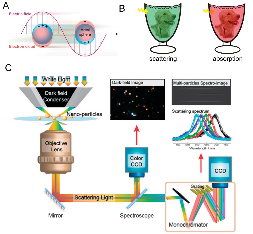
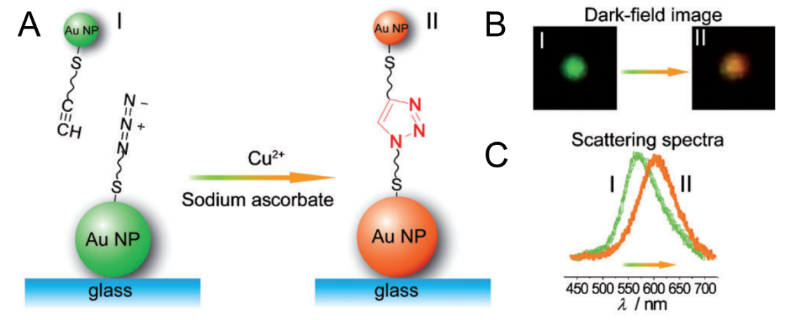
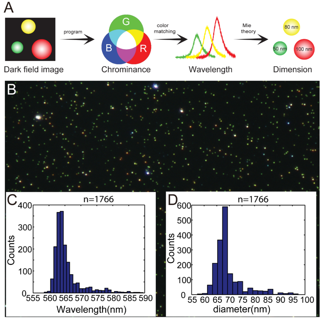
具有独特局域表面等离子共振散射特性的贵金属纳米粒子,在可见光区域表现出明显的吸收和散射光谱特性。在过去的几十年中,基于纳米金和纳米银溶液的可视化颜色传感器,被广泛应用在金属离子、生物分子、农药等灵敏检测。自2000年,暗场显微镜的出现,实现了纳米尺度下等离子共振散射光谱的精准获取,将传感尺度从传统的实验试管发展到单纳米颗粒界面。单颗粒检测消除了本体溶液中大量纳米粒子产生的平均效应,可提供更加准确的反应信息。纳米粒子的散射光谱主要取决于颗粒的尺寸、形貌、成分以及颗粒间耦合作用等,因此,具有特定散射颜色的单个纳米粒子,可以作为优异的纳米探针。这篇综述聚焦于单颗粒纳米传感,首先介绍了纳米粒子局域表面等离子共振的原理和发展历史。随后,主要讨论了单个贵金属纳米粒子作为颜色编码传感器,在生物分子、环境污染物以及能源等领域的应用,尤其是基于单颗粒的原位纳米光谱电化学传感及其在电催化反应中的应用。例如,利用纳米粒子的溶出和生长过程,精巧地设计了针对不同待测物的纳米探针。另一方面,对单纳米粒子结构演变过程的原位监测,也有助于对纳米材料制备机理的理解。最后,着重探讨了纳米颜色传感器信号提取放大的检测手段,包括将肉眼识别的颜色转换为可读的三原色信息以及偏振光检测技术等,进一步扩展单颗粒颜色传感的应用范围。

关键词: 等离子共振散射, 暗场显微镜, 颜色传感器, 纳米电化学, 单颗粒检测

Abstract:

Plasmonic nanoparticles such as Au and Ag with localized surface plasmon resonance (LSPR) property exhibit unique scattering and absorption features. The plasmonic scattering and absorption bands are mainly located at visible light region which can be easily applied in visual detections. By modulating the size, shape and composition of gold and silver colloid solutions, plenty of colorimetric methods have been designed for the detection of metal ions, biomolecules and environmental contaminants. For many years, the LSPR-based measurements are implemented in reagent tubes. Since 2000, the plasmon resonance scattering (PRS) light of metal nanoparticles captured by dark-field microscopy enables the investigation at the nanoscale dimension. Mono-dispersed nanoparticles under a dark-field microscope showed distinct scattering light spots, like colorful stars in the dark sky. The PRS light of a single nanoparticle opens a new way for ultra-sensitive sensing which eliminates the average effects in bulk and provides more accurate reaction information. Thus, individual nanoparticles with specific scattering colors are excellent nanoprobes in the applications of biology, physics, and chemistry. In this review, the plasmonics based colorimetric nanosensors are presented. Particularly, the application of in-situ PRS in the dynamically monitoring of electrocatalytic reactions is highlighted. We firstly introduce a short history of the discovery and development of plasmonic nanoparticles from the ancient artwork to the modern characterization techniques. Some factors including morphology, and dielectric constants that are correlated to the LSPR bands and scattering light colors are listed. Secondly, we demonstrate the use of single plasmonic nanoparticles as visualized color-coded nanoprobes. As the morphology of particles has strong effect on the PRS light, elegant sensors have been conceived by the etching and growth of nanoparticles with different sizes and shapes. On the other hand, the real-time monitoring of particle structure evolution could also reveal the mechanism of the material fabrication at the nanoscale. In addition, core-satellite nanostructures with various linkers are proposed as ultra-sensitive sensors according to the inter-particle coupling effect. Subsequently, we summarize several advanced techniques for nanoscale signal extraction and amplifications. For instance, to expand the application of colorimetric nanosensors, converting the colors into RGB values could clearly distinguish the subtle color changes. Combining with high-throughput signal processing method, thousands of nanoparticles can be rapidly analyzed, which can greatly enhance the measurement efficiency. Except the PRS color, the PRS intensity could also provide abundant information and is easier to be captured. A facile method by converting the PRS intensity of single nanoparticles into visible colors is presented, which is mighty suitable for the in-situ monitoring of fast electrochemical process with high time resolution.

Key words: Plasmon resonance scattering, Dark-field microscopy, Color-coded sensor, Nanoscale electrochemistry, Single-nanoparticle detection