欢迎访问《电化学(中英文)》期刊官方网站,今天是
综述

暗场显微镜下的彩色“纳米星”

  • 静超 ,
  • 龙亿涛
展开
  • a中国科学院上海应用物理研究所,上海 201800
    b生命分析化学国家重点实验室,南京大学化学化工学院,江苏 南京 210023
    c分子传感与成像中心,南京大学,江苏 南京 210023

收稿日期: 2022-12-14

  修回日期: 2023-02-09

  录用日期: 2023-02-20

  网络出版日期: 2023-02-27

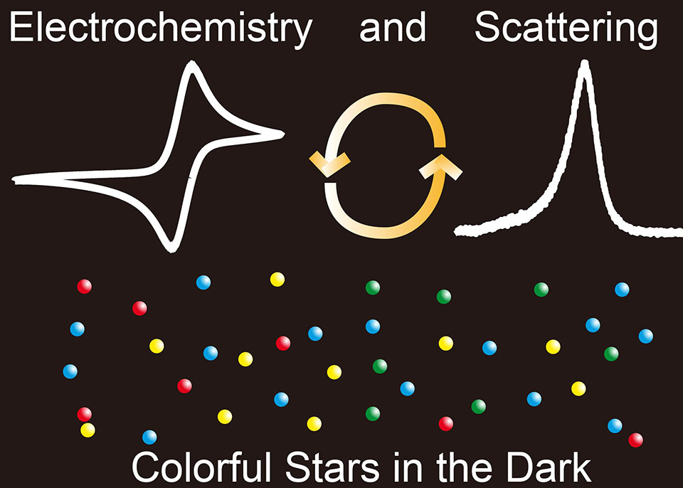
Colorful “Stars” in the Dark

  • Chao Jing ,
  • Yi-Tao Long
Expand
  • aShanghai Institute of Applied Physics, Chinese Academy of Sciences, Shanghai 201800, P.R. China
    bState Key Laboratory of Analytical Chemistry for Life Science; School of Chemistry and Chemical Engineering, Nanjing University, Nanjing 210023, P. R. China
    cMolecular Sensing and Imaging Center, Nanjing University, Nanjing, 210023, PR China
*Tel: (86)13761159439, E-mail: yitaolong@nju.edu.cn

Received date: 2022-12-14

  Revised date: 2023-02-09

  Accepted date: 2023-02-20

  Online published: 2023-02-27

摘要

具有独特局域表面等离子共振散射特性的贵金属纳米粒子,在可见光区域表现出明显的吸收和散射光谱特性。在过去的几十年中,基于纳米金和纳米银溶液的可视化颜色传感器,被广泛应用在金属离子、生物分子、农药等灵敏检测。自2000年,暗场显微镜的出现,实现了纳米尺度下等离子共振散射光谱的精准获取,将传感尺度从传统的实验试管发展到单纳米颗粒界面。单颗粒检测消除了本体溶液中大量纳米粒子产生的平均效应,可提供更加准确的反应信息。纳米粒子的散射光谱主要取决于颗粒的尺寸、形貌、成分以及颗粒间耦合作用等,因此,具有特定散射颜色的单个纳米粒子,可以作为优异的纳米探针。这篇综述聚焦于单颗粒纳米传感,首先介绍了纳米粒子局域表面等离子共振的原理和发展历史。随后,主要讨论了单个贵金属纳米粒子作为颜色编码传感器,在生物分子、环境污染物以及能源等领域的应用,尤其是基于单颗粒的原位纳米光谱电化学传感及其在电催化反应中的应用。例如,利用纳米粒子的溶出和生长过程,精巧地设计了针对不同待测物的纳米探针。另一方面,对单纳米粒子结构演变过程的原位监测,也有助于对纳米材料制备机理的理解。最后,着重探讨了纳米颜色传感器信号提取放大的检测手段,包括将肉眼识别的颜色转换为可读的三原色信息以及偏振光检测技术等,进一步扩展单颗粒颜色传感的应用范围。

本文引用格式

静超 , 龙亿涛 . 暗场显微镜下的彩色“纳米星”[J]. 电化学, 2023 , 29(6) : 2218006 . DOI: 10.13208/j.electrochem.2218006

Abstract

Plasmonic nanoparticles such as Au and Ag with localized surface plasmon resonance (LSPR) property exhibit unique scattering and absorption features. The plasmonic scattering and absorption bands are mainly located at visible light region which can be easily applied in visual detections. By modulating the size, shape and composition of gold and silver colloid solutions, plenty of colorimetric methods have been designed for the detection of metal ions, biomolecules and environmental contaminants. For many years, the LSPR-based measurements are implemented in reagent tubes. Since 2000, the plasmon resonance scattering (PRS) light of metal nanoparticles captured by dark-field microscopy enables the investigation at the nanoscale dimension. Mono-dispersed nanoparticles under a dark-field microscope showed distinct scattering light spots, like colorful stars in the dark sky. The PRS light of a single nanoparticle opens a new way for ultra-sensitive sensing which eliminates the average effects in bulk and provides more accurate reaction information. Thus, individual nanoparticles with specific scattering colors are excellent nanoprobes in the applications of biology, physics, and chemistry. In this review, the plasmonics based colorimetric nanosensors are presented. Particularly, the application of in-situ PRS in the dynamically monitoring of electrocatalytic reactions is highlighted. We firstly introduce a short history of the discovery and development of plasmonic nanoparticles from the ancient artwork to the modern characterization techniques. Some factors including morphology, and dielectric constants that are correlated to the LSPR bands and scattering light colors are listed. Secondly, we demonstrate the use of single plasmonic nanoparticles as visualized color-coded nanoprobes. As the morphology of particles has strong effect on the PRS light, elegant sensors have been conceived by the etching and growth of nanoparticles with different sizes and shapes. On the other hand, the real-time monitoring of particle structure evolution could also reveal the mechanism of the material fabrication at the nanoscale. In addition, core-satellite nanostructures with various linkers are proposed as ultra-sensitive sensors according to the inter-particle coupling effect. Subsequently, we summarize several advanced techniques for nanoscale signal extraction and amplifications. For instance, to expand the application of colorimetric nanosensors, converting the colors into RGB values could clearly distinguish the subtle color changes. Combining with high-throughput signal processing method, thousands of nanoparticles can be rapidly analyzed, which can greatly enhance the measurement efficiency. Except the PRS color, the PRS intensity could also provide abundant information and is easier to be captured. A facile method by converting the PRS intensity of single nanoparticles into visible colors is presented, which is mighty suitable for the in-situ monitoring of fast electrochemical process with high time resolution.

参考文献

[1] Kuwata H, Tamaru H, Esumi K, Miyano K. Resonant light scattering from metal nanoparticles: Practical analysis beyond rayleigh approximation[J]. Appl. Phys. Lett., 2003, 83(22): 4625.
[2] Homola J, Yee S S, Gauglitz G. Surface plasmon resonance sensors: Review[J]. Sens. Actuat. B Chem., 1999, 54(1-2): 3-15.
[3] Eustis S, El-Sayed M A. Why gold nanoparticles are more precious than pretty gold: Noble metal surface plasmon resonance and its enhancement of the radiative and nonradiative properties of nanocrystals of different shapes[J]. Chem. Soc. Rev., 2006, 35(3): 209-217.
[4] Jing C, Reichert J. Nanoscale electrochemistry in the “dark-field”[J]. Curr. Opin. Electrochem., 2017, 6(1): 10-16.
[5] Hu M, Novo C, Funston A, Wang H, Staleva H, Zou S, Mulvaney P, Xia Y, Hartland G V. Dark-field microscopy studies of single metal nanoparticles: Understanding the factors that influence the linewidth of the localized surface plasmon resonance[J]. J. Mater. Chem., 2008, 18(17): 1949-1960.
[6] Anker J N, Hall W P, Lyandres O, Shah N C, Zhao J, Van Duyne R P. Biosensing with plasmonic nanosensors[J]. Nat. Mater., 2008, 7(6): 442-453.
[7] Ma Y, Highsmith A L, Hill C M, Pan S. Dark-Field scattering spectroelectrochemistry analysis of hydrazine oxidation at Au nanoparticle-modified transparent electrodes[J]. J. Phys. Chem. C., 2018, 122(32): 18603-18614.
[8] Wonner K, Evers M V, Tschulik K. Simultaneous opto- and spectro-electrochemistry: Reactions of individual nanoparticles uncovered by dark-field microscopy[J]. J. Am. Chem. Soc., 2018, 140(40): 12658-12661.
[9] Wang H H, He T, Du Y, Wang W H, Shen Y B, Li S P, Zhou X C, Yang F. Evolution of single nanobubbles through multi-state dynamics[J]. Chin. Chem. Lett., 2020, 31(9): 2442-2446.
[10] Wang Y X, Shan X N, Tao N J. Emerging tools for studying single entity electrochemistry[J]. Faraday Discuss., 2016, 193: 9-39.
[11] Oja S M, Wood M, Zhang B. Nanoscale electrochemistry[J]. Anal. Chem., 2013, 85(2): 473-486.
[12] Novo C, Funston A M, Gooding A K, Mulvaney P. Electrochemical charging of single gold nanorods[J]. J. Am. Chem. Soc., 2009, 131(41): 14664-14666.
[13] Jing C, Long Y T. Observing electrochemistry on single plasmonic nanoparticles[J]. Electrochem. Sci. Adv., 2021, 2(4): e2100115.
[14] Shang J, Fan J S, Qin W W, Li K. Single-particle measurements of nanocatalysis with dark-field microscopy[J]. Catalysts, 2022, 12(7): 764.
[15] Olson J, Dominguez-Medina S, Hoggard A, Wang L Y, Chang W S, Link S. Optical characterization of single plasmonic nanoparticles[J]. Chem. Soc. Rev., 2015, 44(1): 40-57.
[16] Wang H H, Zhang T, Zhou X C. Dark-Field spectroscopy: Development, applications and perspectives in single nanoparticle catalysis[J]. J. Phys.: Condens. Matter, 2019, 31(47): 473001.
[17] Asiala S M, Marr J M, Gervinskas G, Juodkazis S, Schultz Z D. Plasmonic color analysis of Ag-coated black-Si SERS substrate[J]. Phys. Chem. Chem. Phys., 2015, 17(45): 30461-30467.
[18] Rodriguez-Fajardo V, Sanz V, de Miguel I, Berthelot J, Acimovic S S, Porcar-Guezenec R, Quidant R. Two-color dark-field (TCDF) microscopy for metal nanoparticle imaging inside cells[J]. Nanoscale, 2018, 10(8): 4019-4027.
[19] Alberti G, Zanoni C, Magnaghi L R, Biesuz R. Gold and silver nanoparticle-based colorimetric sensors: New trends and applications[J]. Chemosensors, 2021, 9(11): 305.
[20] Wang S M, Wang H, Zhao W, Xu J J, Chen H Y. Single-particle detection of cholesterol based on the host-guest recognition induced plasmon resonance energy transfer[J]. Chin. Chem. Lett., 2022: 108053.
[21] Liu G Y, Lu M, Huang X D, Li T F, Xu D H. Application of gold-nanoparticle colorimetric sensing to rapid food safety screening[J]. Sensors, 2018, 18(12): 4166.
[22] Sharma R, Dhillon A, Kumar D. Mentha-stabilized silver nanoparticles for high-performance colorimetric detection of Al(III) in aqueous systems[J]. Sci. Rep., 2018, 8(1): 5189.
[23] Li Y, Jing C, Zhang L, Long Y T. Resonance scattering particles as biological nanosensors in vitro and in vivo[J]. Chem. Soc. Rev., 2012, 41(2): 632-642.
[24] Hafner J H, Mayer K M. Localized surface plasmon resonance sensors[J]. Chem. Rev., 2011, 111(6): 3828-3857.
[25] Kelly K L, Coronado E, Zhao L L, Schatz G C. The optical properties of metal nanoparticles: The influence of size, shape, and dielectric environment[J]. J. Phys. Chem. B, 2003, 107(3): 668-677.
[26] Stewart M E, Anderton C R, Thompson L B, Maria J, Gray S K, Rogers J A, Nuzzo R G. Nanostructured plasmonic sensors[J]. Chem. Rev., 2008, 108(2): 494-521.
[27] Sonnichsen C, Geier S, Hecker N E, von Plessen G, Feldmann J, Ditlbacher H, Lamprecht B, Krenn J R, Aussenegg F R, Chan V Z H, Spatz J P, Moller M. Spectroscopy of single metallic nanoparticles using total internal reflection microscopy[J]. Appl. Phys. Lett., 2000, 77(19): 2949-2951.
[28] Schultz D A, Schultz S, Smith D R, Mock J J. Single-target molecule detection with nonbleaching multicolor optical immunolabels[J]. Proc. Natl. Acad. Sci., 2000, 97(3): 996-1001.
[29] Nehl C L, Liao H, Hafner J H. Optical properties of star-shaped gold nanoparticles[J]. Nano Lett., 2006, 6(4): 683-688.
[30] Jain P K, El-Sayed I H, El-Sayed M A. Au nanoparticles target cancer[J]. Nano Today, 2007, 2(1): 18-29.
[31] Mock J J, Barbic M, Smith D R, Schultz D A, Schultz S. Shape effects in plasmon resonance of individual colloidal silver nanoparticles[J]. J. Chem. Phys., 2002, 116(15): 6755-6759.
[32] Liu Y, Ling J, Huang C Z. Individually color-coded plasmonic nanoparticles for rgb analysis[J]. Chem. Commun., 2011, 47(28): 8121-8123.
[33] Hill C M, Pan S. A Dark-Field scattering spectroelectrochemical technique for tracking the electrodeposition of single silver nanoparticles[J]. J. Am. Chem. Soc., 2013, 135(46): 17250-17253.
[34] Qin L X, Li Y, Li D W, Jing C, Chen B Q, Ma W, Heyman A, Shoseyov O, Willner I, Tian H, Long Y T. Electrodeposition of single-metal nanoparticles on stable protein 1 membranes: application of plasmonic sensing by single nanoparticles[J]. Angew. Chem. Int. Ed., 2012, 51(1): 140-144.
[35] Hill C M, Bennett R, Zhou C, Street S, Zheng J, Pan S. Single Ag nanoparticle spectroelectrochemistry via dark-field scattering and fluorescence microscopies[J]. J. Phys. Chem. C., 2015, 119(12): 6760-6768.
[36] Wonner K, Rurainsky C, Tschulik K. Operando studies of the electrochemical dissolution of silver nanoparticles in nitrate solutions observed with hyperspectral dark-field microscopy[J]. Front Chem., 2019, 7: 912.
[37] Sun S S, Gao M X, Lei G, Zou H Y, Ma J, Huang C Z. Visually monitoring the etching process of gold nanoparticles by KI/I2 at single-nanoparticle level using scattered-light dark-field microscopic imaging[J]. Nano Res., 2016, 9(4): 1125-1134.
[38] Hu S, Yi J, Zhang Y J, Lin K Q, Liu B J, Chen L, Zhan C, Lei Z C, Sun J J, Zong C, Li J F, Ren B. Observing atomic layer electrodeposition on single nanocrystals surface by dark field spectroscopy[J]. Nat. Commun., 2020, 11(1): 2518.
[39] Qin L X, Jing C, Li Y, Li D W, Long Y T. Real-time monitoring of the aging of single plasmonic copper nanoparticles[J]. Chem. Commun., 2012, 48(10): 1511-1513.
[40] Chirea M, Collins S S, Wei X, Mulvaney P. Spectroelectrochemistry of silver deposition on single gold nanocrystals[J]. J. Phys. Chem. Lett., 2014, 5(24): 4331-4335.
[41] Hwang C S H, Ahn M S, Lee Y, Chung T, Jeong K H. Ag/Au alloyed nanoislands for wafer-level plasmonic color filter arrays[J]. Sci. Rep., 2019, 9(1): 9082.
[42] Wang J G, Fossey J S, Li M, Xie T, Long Y T. Real-time plasmonic monitoring of single gold amalgam nanoalloy electrochemical formation and stripping[J]. ACS Appl. Mater. Interface, 2016, 8(12): 8305-8314.
[43] Liu Y, Huang C Z. Real-time dark-field scattering microscopic monitoring of the in situ growth of single Ag@Hg nanoalloys[J]. ACS Nano, 2013, 7(12): 11026-11034.
[44] Wang H, Zhao W, Xu C H, Chen H Y, Xu J J. Electrochemical synthesis of Au@semiconductor core-shell nanocrystals guided by single particle plasmonic imaging[J]. Chem. Sci., 2019, 10(40): 9308-9314.
[45] Liu Q, Jing C, Zheng X, Gu Z, Li D, Li D W, Huang Q, Long Y T, Fan C. Nanoplasmonic detection of adenosine triphosphate by aptamer regulated self-catalytic growth of single gold nanoparticles[J]. Chem. Commun., 2012, 48(77): 9574-9576.
[46] Gu X Y, Liu J J, Gao P F, Li Y F, Huang C Z. Gold triangular nanoplates based single-particle dark-field microscopy assay of pyrophosphate[J]. Anal. Chem., 2019, 91(24): 15798-15803.
[47] Ye Z J, Weng R, Ma Y H, Wang F Y, Liu H, Wei L, Xiao L H. Label-free, single-particle, colorimetric detection of permanganate by GNPs@Ag core-shell nanoparticles with dark-field optical microscopy[J]. Anal. Chem., 2018, 90(21): 13044-13050.
[48] Huang M N, Fan Y P, Yuan X, Wei L. Color-coded detection of malathion based on enzyme inhibition with dark-field optical microscopy[J]. Sens. Actuat. B Chem., 2022, 353(15): 131135.
[49] Zhang L, Li Y, Li D W, Jing C, Chen X, Lv M, Huang Q, Long Y T, Willner I. Single gold nanoparticles as real-time optical probes for the detection of NADH-dependent intracellular metabolic enzymatic pathways[J]. Angew. Chem. Int. Ed., 2011, 50(30): 6789-6792.
[50] Qi F, Han Y M, Ye Z J, Liu H, Wei L, Xiao L H. Color-coded single-particle pyrophosphate assay with dark-field optical microscopy[J]. Anal. Chem., 2018, 90(18): 11146-11153.
[51] Pini V, Kosaka P M, Ruz J J, Malvar O, Encinar M, Tamayo J, Calleja M. Spatially multiplexed dark-field microspectrophotometry for nanoplasmonics[J]. Sci. Rep., 2016, 6: 22836.
[52] Ghosh S K, Pal T. Interparticle coupling effect on the surface plasmon resonance of gold nanoparticles:? From theory to applications[J]. Chem. Rev., 2007, 107(11): 4797-4862.
[53] Sonnichsen C, Reinhard B M, Liphardt J, Alivisatos A P. A molecular ruler based on plasmon coupling of single gold and silver nanoparticles[J]. Nat. Biotechnol., 2005, 23(6): 741-745.
[54] Xiao L, Wei L, He Y, Yeung E S. Single molecule biosensing using color coded plasmon resonant metal nanoparticles[J]. Anal. Chem., 2010, 82(14): 6308-6314.
[55] Jin H Y, Li D W, Zhang N, Gu Z, Long Y T. Analyzing carbohydrate-protein interaction based on single plasmonic nanoparticle by conventional Dark Field microscopy[J]. ACS Appl. Mater. Interface, 2015, 7(22): 12249-12253.
[56] Shi L, Jing C, Ma W, Li D W, Halls J E, Marken F, Long Y T. Plasmon resonance scattering spectroscopy at the single-nanoparticle level: Real-time monitoring of a click reaction[J]. Angew. Chem. Int. Ed., 2013, 52(23): 6011-6014.
[57] Ding T, Mertens J, Lombardi A, Scherman O A, Baumberg J J. Light-directed tuning of plasmon resonances via plasmon-induced polymerization using hot electrons[J]. ACS Photonics, 2017, 4(6): 1453-1458.
[58] Hao J, Xiong B, Cheng X, He Y, Yeung E S. High-throughput sulfide sensing with colorimetric analysis of single Au-Ag core-shell nanoparticles[J]. Anal. Chem., 2014, 86(10): 4663-4667.
[59] Zhou J, Yang T, He W, Pan Z Y, Huang C Z. A galvanic exchange process visualized on single silver nanoparticles via dark-field microscopy imaging[J]. Nanoscale, 2018, 10(26): 12805-12812.
[60] Aaron J, Travis K, Harrison N, Sokolov K. Dynamic imaging of molecular assemblies in live cells based on nanoparticle plasmon resonance coupling[J]. Nano Lett., 2009, 9(10): 3612-3618.
[61] Gu Z, Jing C, Ying Y L, He P, Long Y T. In situ high throughput scattering light analysis of single plasmonic nanoparticles in living cells[J]. Theranostics, 2015, 5(2): 188-195.
[62] Zhou J, Lei G, Zheng L L, Gao P F, Huang C Z. Hsi colour-coded analysis of scattered light of single plasmonic nanoparticles[J]. Nanoscale, 2016, 8(22): 11467-11471.
[63] Zhou J, Gao P F, Zhang H Z, Lei G, Zheng L L, Liu H, Huang C Z. Color resolution improvement of the dark-field microscopy imaging of single light scattering plasmonic nanoprobes for microrna visual detection[J]. Nanoscale, 2017, 9(13): 4593-4600.
[64] Sriram M, Markhali B P, Nicovich P R, Bennett D T, Reece P J, Brynn Hibbert D, Tilley R D, Gaus K, Vivekchand S R C, Gooding J J. A rapid readout for many single plasmonic nanoparticles using dark-field microscopy and digital color analysis[J]. Biosens. Bioelectron., 2018, 117: 530-536.
[65] Jing C, Gu Z, Ying Y L, Li D W, Zhang L, Long Y T. Chrominance to dimension: A real-time method for measuring the size of single gold nanoparticles[J]. Anal. Chem., 2012, 84(10): 4284-4291.
[66] Wagner T, Lipinski H G, Wiemann M. Dark Field nanoparticle tracking analysis for size characterization of plasmonic and non-plasmonic particles[J]. J. Nanopart. Res., 2014, 16(5): 2419.
[67] Huang Y, Kim D H. Dark-Field microscopy studies of polarization-dependent plasmonic resonance of single gold nanorods: rainbow nanoparticles[J]. Nanoscale, 2011, 3(8): 3228-3232.
[68] Liu J J, Yan H H, Zhang Q, Gao P F, Li C M, Liang G L, Huang C Z, Wang J. High-resolution vertical polarization excited dark-field microscopic imaging of anisotropic gold nanorods for the sensitive detection and spatial imaging of intracellular microrna-21[J]. Anal. Chem., 2020, 92(19): 13118-13125.
[69] Fan J R, Wu W G, Chen Z J, Zhu J, Li J. Three-dimensional cavity nanoantennas with resonant-enhanced surface plasmons as dynamic color-tuning reflectors[J]. Nanoscale, 2017, 9(10): 3416-3423.
[70] Ng R J H, Krishnan R V, Wang H, Yang J K W. Darkfield colors from multi-periodic arrays of gap plasmon resonators[J]. Nanophotonics, 2020, 9(2): 533-545.
[71] Wang J G, Fossey J S, Li M, Li D W, Ma W, Ying Y L, Qian R C, Cao C, Long Y T. Real-time plasmonic monitoring of electrocatalysis on single nanorods[J]. J. Electroanal. Chem., 2016, 781: 257-264.
[72] Zhou H, Liu Q, Rawson F J, Ma W, Li D W, Li D, Long YT. Optical monitoring of faradaic reaction using single plasmon-resonant nanorods functionalized with graphene[J]. Chem. Commun., 2015, 51(15): 3223-3226.
[73] Cao Y, Zhou H, Qian R C, Liu J, Ying Y L, Long Y T. Analysis of the electron transfer properties of carbon quantum dots on gold nanorod surfaces via plasmonic resonance scattering spectroscopy[J]. Chem. Commun., 2017, 53(42): 5729-5732.
[74] Jing C, Gu Z, Long Y T. Imaging electrocatalytic processes on single gold nanorods[J]. Faraday Discuss., 2016, 193: 371-385.
[75] Jing C, Gu Z, Xie T, Long Y T. Color-coded imaging of electrochromic process at single nanoparticle level[J]. Chem. Sci., 2016, 7(8): 5347-5351.
文章导航

/