
电化学(中英文) ›› 2021, Vol. 27 ›› Issue (2): 208-215. doi: 10.13208/j.electrochem.201245
吴丽文1, 王玮2, 黄逸凡1,*( )
)
收稿日期:2021-01-29
修回日期:2021-03-18
出版日期:2021-04-28
发布日期:2021-03-20
通讯作者:
黄逸凡
E-mail:huangyf@shanghaitech.edu.cn
基金资助:
Li-Wen Wu1, Wei Wang2, Yi-Fan Huang1,*()
Received:2021-01-29
Revised:2021-03-18
Published:2021-04-28
Online:2021-03-20
Contact:
Yi-Fan Huang
E-mail:huangyf@shanghaitech.edu.cn
摘要:
镍(Ni)电极在电化学中应用广泛。原位表征Ni电极表面的吸附物种有益于帮助理解电极反应历程、指导发展高效电催化剂。应用超微电极作为工作电极的电化学表面增强拉曼光谱技术结合了超微电极表面的传质特性和分子水平的高灵敏度表征,是研究Ni电化学的有力手段。本文所述的研究工作通过在金(Au)超微电极表面电吸附具有SERS活性的Au纳米粒子并恒电流沉积金属Ni薄层,制备并表征了具有SERS活性的Ni超微电极。在氢氧化钠溶液中的循环伏安实验和以4-甲基苯硫酚分子作为探针分子的SERS实验结果表明,沉积速率和沉积电量是影响超微电极表面Ni的覆盖度和SERS活性的关键因素。在吸附了直径为55 nm Au纳米粒子的、直径为10 μm Au的超微电极表面,以100 μA·cm-2电流密度电沉积厚度约为5个原子层Ni的条件下,可获得Ni覆盖完好的、具有最强SERS活性的Ni超微电极。
吴丽文, 王玮, 黄逸凡. 应用镍超微电极的电化学表面增强拉曼光谱技术研究[J]. 电化学(中英文), 2021, 27(2): 208-215.
Li-Wen Wu, Wei Wang, Yi-Fan Huang. Electrochemical Surface-Enhanced Raman Spectroscopic Studies on Nickel Ultramicroelectrode[J]. Journal of Electrochemistry, 2021, 27(2): 208-215.

图1
55 nm Au纳米粒子的扫描电镜图
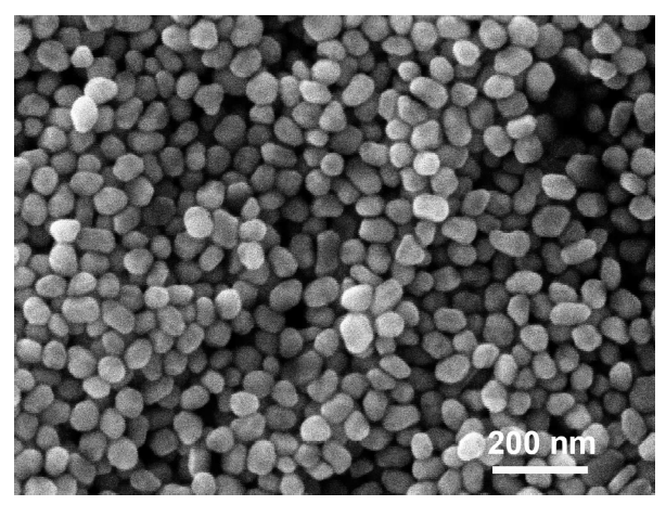
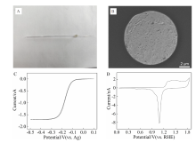
图2
A. 直径10 μm的Au超微电极的照片;B. 直径10 μm的Au超微电极的扫描电镜图;C. Au超微电极在1 mmol·L-1 Ru(NH3)6Cl3 + 0.1 mol·L-1 KCl溶液中的循环伏安曲线,扫速为10 mV·s-1;D. Au超微电极在0.1 mol·L-1 HClO4溶液中的循环伏安曲线,扫速为500 mV·s-1。


图3
A. 吸附Au纳米粒子的Au超微电极的扫描电镜图;B. Au超微电极吸附纳米粒子前/后在0.1 mol·L-1 HClO4 溶液中的循环伏安曲线,扫速为500 mV·s-1。
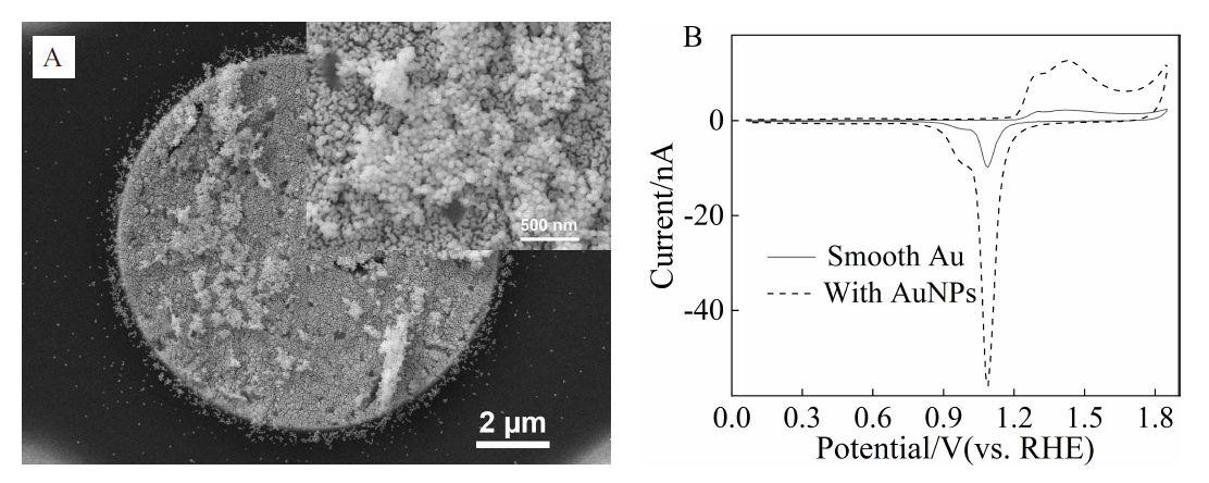
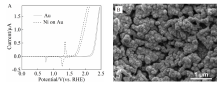
图4
A. 沉积了Ni的Au超微电极在0.1 mol·L-1 NaOH中的循环伏安曲线,扫速为500 mV·s-1;B. 沉积了Ni的Au超微电极的SEM图。

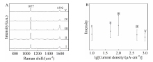
图5
A. 4-甲基苯硫酚吸附在以不同电流密度沉积形成的Ni超微电极上的SERS谱图: I. 10 μA·cm-2, II. 50 μA·cm-2, III. 100 μA·cm-2, IV. 500 μA·cm-2, V. 1000 μA·cm-2; B. 电沉积电流密度与1077 cm-1强度关系图。


图6
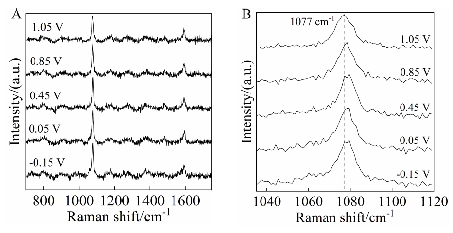
A. 吸附4-甲基苯硫酚的Ni超微电极在0.1 mol·L-1 NaOH溶液中不同电位时的SERS谱图;B. 不同电极电势下频率为1077 cm-1的SERS峰。
| [1] |
Ni W Y, Krammer A, Hsu C S, Chen H M, Schuler A, Hu X L. Ni3N as an active hydrogen oxidation reaction catalyst in alkaline medium[J]. Angew. Chem. In. Ed., 2019,58(22):7445-7449.
doi: 10.1002/anie.v58.22 URL |
| [2] | Lu X F, Yu L, Lou X W. Highly crystalline Ni-doped FeP/carbon hollow nanorods as all-pH efficient and durable hydrogen evolving electrocatalysts[J]. Sci. Adv., 2019, 5(2): eaav6009. |
| [3] |
Wang Q, Huang X, Zhao Z L, Wang M Y, Xiang B, Li J, Feng Z X, Xu H, Gu M. Ultrahigh-loading of Ir single atoms on NiO matrix to dramatically enhance oxygen evolution reaction[J]. J. Am. Chem. Soc., 2020,142(16):7425-7433.
doi: 10.1021/jacs.9b12642 URL |
| [4] |
Stamenkovic V R, Fowler B, Mun B S, Wang G F, Ross P N, Lucas C A, Markovic N M. Improved oxygen reduction activity on Pt3Ni(111) via increased surface site availability[J]. Science, 2007,315(5811):493-497.
doi: 10.1126/science.1135941 URL |
| [5] |
Yang H B, Hung S F, Liu S, Yuan K D, Miao S, Zhang L P, Huang X, Wang H Y, Cai W Z, Chen R, Gao J J, Yang X F, Chen W, Huang Y Q, Chen H M, Li C M, Zhang T, Liu B. Atomically dispersed Ni(I) as the active site for electrochemical CO2 reduction[J]. Nat. Energy, 2018,3(2):140-147.
doi: 10.1038/s41560-017-0078-8 URL |
| [6] |
Wang H L, Casalongue H S, Liang Y Y, Dai H J. Ni(OH)2 nanoplates grown on graphene as advanced electrochemical pseudocapacitor materials[J]. J. Am. Chem. Soc., 2010,132(21):7472-7477.
doi: 10.1021/ja102267j URL |
| [7] |
Sun H C, Qin D, Huang S Q, Guo X Z, Li D M, Luo Y H, Meng Q B. Dye-sensitized solar cells with NiS counter electrodes electrodeposited by a potential reversal technique[J]. Energy Environ. Sci., 2011,4(8):2630-2637.
doi: 10.1039/c0ee00791a URL |
| [8] | Bard A J, Faulkner L R. Electrochemical methods: fundamentals and applications[M]. New York: John Wiley & Sons, Inc., 2001: 168-176. |
| [9] |
Fleischmann M, Hendra P J, McQuillan A J. Raman spectra of pyridine adsorbed at a silver electrode[J]. Chem. Phys. Lett., 1974,26(2):163-166.
doi: 10.1016/0009-2614(74)85388-1 URL |
| [10] |
Albrecht M G, Creighton J A. Anomalously intense Raman spectra of pyridine at a silver electrode[J]. J. Am. Chem. Soc., 1977,99(15):5215-5217.
doi: 10.1021/ja00457a071 URL |
| [11] |
Jeanmaire D L, Van Duyne R P. Surface raman spectroelectrochemistry: Part I. Heterocyclic, aromatic, and aliphatic amines adsorbed on the anodized silver electrode[J]. J. Electroanal. Chem., 1977,84(1):1-20.
doi: 10.1016/S0022-0728(77)80224-6 URL |
| [12] |
Wu D Y, Li J F, Ren B, Tian Z Q. Electrochemical surface-enhanced Raman spectroscopy of nanostructures[J]. Chem. Soc. Rev., 2008,37(5):1025-1041.
doi: 10.1039/b707872m URL |
| [13] |
Kostecki R, McLarnon F. Electrochemical and in situ Raman spectroscopic characterization of nickel hydroxide electrodes: I. Pure nickel hydroxide[J]. J. Electrochem. Soc., 1997,144:485-493.
doi: 10.1149/1.1837437 URL |
| [14] |
Diaz-Morales O, Ferrus-Suspedra D, Koper M T M. The importance of nickel oxyhydroxide deprotonation on its activity towards electrochemical water oxidation[J]. Chem. Sci., 2016,7(4):2639-2645.
doi: 10.1039/c5sc04486c pmid: 28660036 |
| [15] |
Louie M W, Bell A T. An investigation of thin-film Ni-Fe oxide catalysts for the electrochemical evolution of oxygen[J]. J. Am. Chem. Soc., 2013,135(33):12329-12337.
doi: 10.1021/ja405351s URL |
| [16] |
Wang Y H, Wang X T, Ze H, Zhang X G, Radjenovic P M, Zhang Y J, Dong J C, Tian Z Q, Li J F. Spectroscopic verification of adsorbed hydroxyl intermediate in the bifunctional mechanism of hydrogen oxidation reaction[J]. Angew. Chem. Int. Ed., 2021,60(11):5708-5711.
doi: 10.1002/anie.v60.11 URL |
| [17] |
Kim B J, Lee D J, Kim Y R, Lim S Y, Bae J H, Kim K B, Chung T D. Gold microshell tip for in situ electrochemical Raman spectroscopy[J]. Adv. Mater., 2012,24(3):421-424.
doi: 10.1002/adma.201103644 URL |
| [18] | Wang W, Huang Y F, Liu D Y, Wang F F, Tian Z Q, Zhan D P. Electrochemically roughened gold microelectrode for surface-enhanced Raman spectroscopy[J]. J. Ele-ctroanal. Chem., 2016,779:126-130. |
| [19] |
Huang Y F, Wang W, Guo H Y, Zhan C, Duan S, Zhan D P, Wu D Y, Ren B, Tian Z Q. Microphotoelectrochemical surface-enhanced Raman spectroscopy: toward bridging hot-electron transfer with a molecular reaction[J]. J. Am. Chem. Soc., 2020,142(18):8483-8489.
doi: 10.1021/jacs.0c02523 URL |
| [20] |
Liu N Y, Wu L W, Huang Y F. In-situ electrochemical Raman spectroscopy on ultramicroelectrodes[J]. Sci. Sin. Chim., 2021,51(3):256-263.
doi: 10.1360/SSC-2020-0179 URL |
| [21] |
Ren B, Huang Q J, Cai W B, Mao B W, Liu F M, Tian Z Q. Surface Raman spectra of pyridine and hydrogen on bare platinum and nickel electrodes[J]. J. Electroanal. Chem., 1996,415(1-2):175-178.
doi: 10.1016/S0022-0728(96)01004-2 URL |
| [22] |
Huang Q J, Yao J L, Mao B W, Gu R A, Tian Z Q. Surface Raman spectroscopic studies of pyrazine adsorbed onto nickel electrodes[J]. Chem. Phys. Lett., 1997,271(1-3):101-106.
doi: 10.1016/S0009-2614(97)00419-3 URL |
| [23] |
Tian Z Q, Ren B, Wu D Y. Surface-enhanced Raman scattering: from noble to transition metals and from rough surfaces to ordered nanostructures[J]. J. Phys. Chem. B, 2002,106(37):9463-9483.
doi: 10.1021/jp0257449 URL |
| [24] | Gao J S(高劲松), Ren B(任斌), Huang Q J(黄群健), Tian Z Q(田中群). Surface Raman spectra obtained from various electrodeposited transidon metals[J]. J. Electro-chem. (电化学) 1996,2(3):258-261. |
| [25] |
Xia Y Y, Wu Y W, Wu L W, Wang T Y Y, Hang T, Huang Y F, Li M. Two-step electrodeposited 3D Ni nanocone supported Au nanoball arrays as SERS substrate[J]. J. Electrochem. Soc., 2020,167(14):142502.
doi: 10.1149/1945-7111/abc0aa URL |
| [26] |
Fleischmann M, Tian Z Q, Li L J. Raman spectroscopy of adsorbates on thin film electrodes deposited on silver substrates[J]. J. Electroanal. Chem., 1987,217(2):397-410.
doi: 10.1016/0022-0728(87)80231-0 URL |
| [27] |
Fleischmann M, Tian Z Q. The induction of SERS on smooth Ag by the deposition of Ni and Co[J]. J. Electro-anal. Chem., 1987,217(2):411-416.
doi: 10.1016/0022-0728(87)80232-2 URL |
| [28] |
Bao F, Li J F, Ren B, Yao J L, Gu R A, Tian Z Q. Synjournal and characterization of Au@Co and Au@Ni core-shell nanoparticles and their applications in surface-enhanced Raman spectroscopy[J]. J. Phys. Chem. C, 2008,112(2):345-350.
doi: 10.1021/jp075844k URL |
| [29] |
Moskovits M. Surface-enhanced spectroscopy[J]. Rev. Mod. Phys., 1985,57(3):783-826.
doi: 10.1103/RevModPhys.57.783 URL |
| [30] | Willets K A, Van Duyne R P. Localized surface plasmon resonance spectroscopy and sensing[M]. Annual Review of Physical Chemistry, 2007,58:267-297. |
| [31] |
Hayazawa N, Inouye Y, Sekkat Z, Kawata S. Metallized tip amplification of near-field Raman scattering[J]. Opt. Commun., 2000,183(1-4):333-336.
doi: 10.1016/S0030-4018(00)00894-4 URL |
| [32] |
Stöckle R M, Suh Y D, Deckert V, Zenobi R. Nanoscale chemical analysis by tip-enhanced Raman spectroscopy[J]. Chem. Phys. Lett., 2000,318(1-3):131-136.
doi: 10.1016/S0009-2614(99)01451-7 URL |
| [33] |
Pettinger B, Ren B, Picardi G, Schuster R, Ertl G. Nano-scale probing of adsorbed species by tip-enhanced Raman spectroscopy[J]. Phys. Rev. Lett., 2004,92(9):096101.
doi: 10.1103/PhysRevLett.92.096101 URL |
| [34] | Tian Z Q, Ren B, Li J F, Yang Z L. Expanding generality of surface-enhanced Raman spectroscopy with borrowing SERS activity strategy[J]. Chem. Commun., 2007: 3514-3534. |
| [35] |
Li J F, Zhang Y J, Ding S Y, Panneerselvam R, Tian Z Q. Core-shell nanoparticle-enhanced Raman spectroscopy[J]. Chem. Rev., 2017,117(7):5002-5069.
doi: 10.1021/acs.chemrev.6b00596 URL |
| [36] |
Montelongo Y, Sikdar D, Ma Y, McIntosh A J S, Velleman L, Kucernak A R, Edel J B, Kornyshev A A. Electrotunable nanoplasmonic liquid mirror[J]. Nat. Mater., 2017,16(11):1127-1135.
doi: 10.1038/nmat4969 pmid: 28892055 |
| [37] |
Frens G. Controlled nucleation for the regulation of the particle size in monodisperse gold suspensions[J]. Nature Phys. Sci., 1973,241(105):20-22.
doi: 10.1038/physci241020a0 URL |
| [38] |
Shao Y, Mirkin M V, Fish G, Kokotov S, Palanker D, Lewis A. Nanometer-sized electrochemical sensors[J]. Anal. Chem., 1997,69(8):1627-1634.
doi: 10.1021/ac960887a URL |
| [39] |
Sun P, Mirkin M V. Kinetics of electron-transfer reactions at nanoelectrodes[J]. Anal. Chem., 2006,78(18):6526-6534.
doi: 10.1021/ac060924q URL |
| [40] |
Zhan D, Velmurugan J, Mirkin M V. Adsorption/desorption of hydrogen on Pt nanoelectrodes: evidence of surface diffusion and spillover[J]. J. Am. Chem. Soc., 2009,131(41):14756-14760.
doi: 10.1021/ja902876v URL |
| [41] |
Ma Y, Sikdar D, Fedosyuk A, Ma Y, Sikdar D, Fedosyuk A, Velleman L, Klemme D J, Oh S H, Kucernak ARJ, Kornyshev A A, Edel J B. Electrotunable nanoplasmonics for amplified surface enhanced Raman spectroscopy[J]. ACS Nano, 2020,14(1):328-336.
doi: 10.1021/acsnano.9b05257 URL |
| [42] |
Nie S, Emory S R. Probing single molecules and single nanoparticles by surface-enhanced Raman scattering[J]. Science, 1997,275(5303):1102-1106.
doi: 10.1126/science.275.5303.1102 URL |
| [43] |
Kneipp K, Wang Y, Kneipp H, Perelman, L T, Itzkan I, Dasari R, Feld M S. Single molecule detection using surface-enhanced Raman scattering (SERS)[J]. Physical Review Letters, 1997,78(9):1667-1670.
doi: 10.1103/PhysRevLett.78.1667 URL |
| [44] |
Xu H X, Aizpurua J, Käll M, Apell P. Electromagnetic contributions to single-molecule sensitivity in surface-enhanced Raman scattering[J]. Phys. Rev. E, 2000,62(3):4318-4324.
doi: 10.1103/PhysRevE.62.4318 URL |
| [1] | 马桢, 林佳阳, 南文静, 韩联欢, 詹东平. 超微电极实验:基本原理、制备方法和伏安性能[J]. 电化学(中英文), 2023, 29(7): 2216002-. |
| [2] | 谭卓, 李凯旋, 毛秉伟, 颜佳伟. 电化学扫描隧道显微术:以Cu在Au(111)表面初始阶段电沉积为例[J]. 电化学(中英文), 2023, 29(7): 2216003-. |
| [3] | 李家欣, 冯立纲. 析氧反应铁镍基预催化剂的表界面调控与进展[J]. 电化学(中英文), 2022, 28(9): 2214001-. |
| [4] | 郭丹丹, 俞红梅, 迟军, 邵志刚. 自支撑NiFe LDHs@Co-OH-CO3纳米棒阵列电极用于碱性阴离子交换膜电解水[J]. 电化学(中英文), 2022, 28(9): 2214003-. |
| [5] | 王京玥, 王睿, 王诗琦, 王立帆, 詹纯. 一步固相法合成锂离子电池高镍层状正极材料[J]. 电化学(中英文), 2022, 28(8): 2112131-. |
| [6] | 杨家强, 金磊, 李威青, 王赵云, 杨防祖, 詹东平, 田中群. 亚硫酸盐无氰电沉积金新工艺及机制[J]. 电化学(中英文), 2022, 28(7): 2213005-. |
| [7] | 孙云娜, 吴永进, 谢东东, 蔡涵, 王艳, 丁桂甫. 硅通孔内铜电沉积填充机理研究进展[J]. 电化学(中英文), 2022, 28(7): 2213001-. |
| [8] | 黄葵, 黄容姣, 刘素琴, 何震. 电子功能外延薄膜的电沉积[J]. 电化学(中英文), 2022, 28(7): 2213006-. |
| [9] | 倪修任, 张雅婷, 王翀, 洪延, 陈苑明, 苏元章, 何为, 陈先明, 黄本霞, 续振林, 李毅峰, 李能彬, 杜永杰. 电沉积纳米锥镍的生长机理及其性能的研究[J]. 电化学(中英文), 2022, 28(7): 2213008-. |
| [10] | 缪桦, 李明瑞, 邹文中, 周国云, 王守绪, 叶晓菁, 朱凯. Sn-Ag-Cu三元合金焊料电沉积中添加剂的影响研究[J]. 电化学(中英文), 2022, 28(6): 2104411-. |
| [11] | 魏丽君, 周紫晗, 吴蕴雯, 李明, 王溯. 芯片钴互连及其超填充电镀技术的研究进展[J]. 电化学(中英文), 2022, 28(6): 2104431-. |
| [12] | 沈银飞, 陈艳丽, 王笙戌, 朱晔, 王文昌, 吴敏娴, 陈智栋. 酸性溶液中苯并三氮唑和3-巯基-1-丙烷磺酸钠在铜电极表面的电化学SERS研究[J]. 电化学(中英文), 2022, 28(6): 2104451-. |
| [13] | 战充波, 张润佳, 付旭, 孙海静, 周欣, 王保杰, 孙杰. 氯离子对ChCl-Urea低共熔溶剂中银电沉积的电化学行为影响[J]. 电化学(中英文), 2022, 28(5): 2111151-. |
| [14] | 王昊, 曹晓舟, 薛向欣. 锑在氯化胆碱-乙二醇低共熔溶剂中的电沉积研究[J]. 电化学(中英文), 2022, 28(4): 2103071-. |
| [15] | 彭辉远, 王家正, 刘佳, 于欢欢, 林建德, 吴德印, 田中群. 纳米结构金电极上对氨基苯硫酚的电化学反应过程研究[J]. 电化学(中英文), 2022, 28(4): 2106281-. |
| 阅读次数 | ||||||
|
全文 |
|
|||||
|
摘要 |
|
|||||